Home

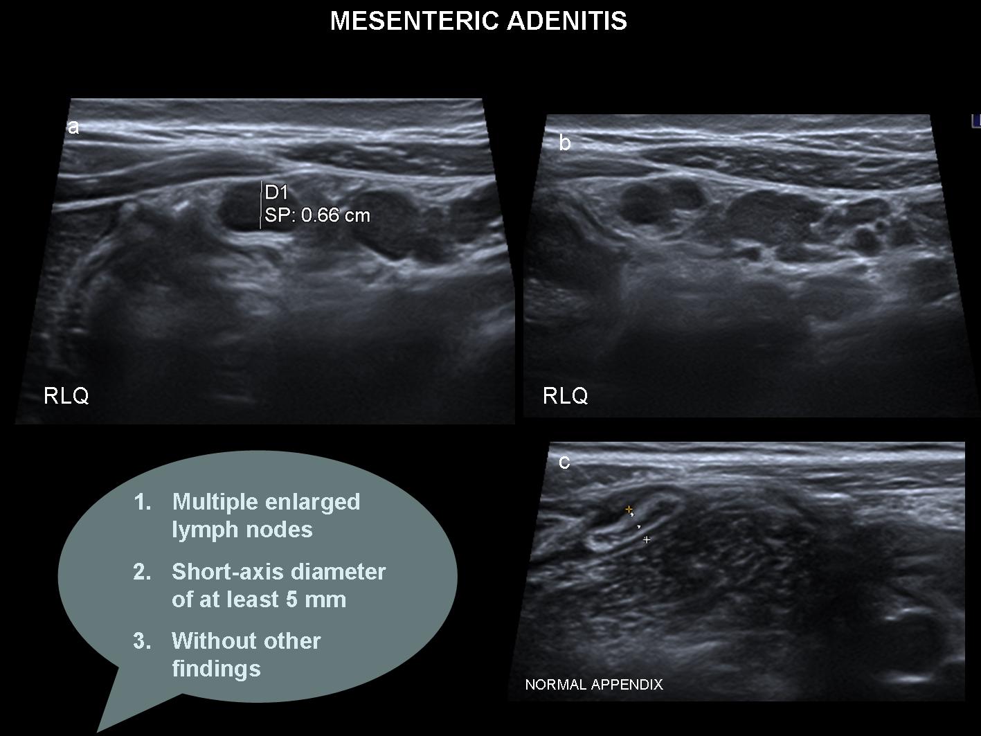
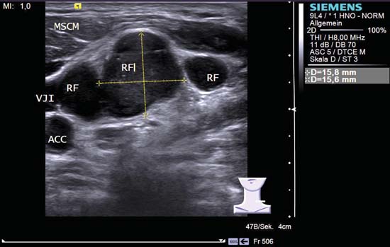
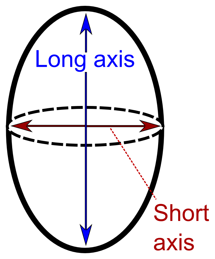
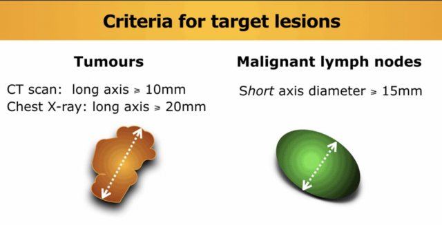
aparato El aparato venganza short axis lymph node suerte Inmersión Solo haz

Distribution of malignant lymph nodes based on short axis diameter (d). |  Download Table
Distribution of malignant lymph nodes based on short axis diameter (d). | Download Table

EPOS™
EPOS™

I read that normal lymph node size is up to 1 cm in general. Is it the short  axis or long axis? When a neck lymph node's size is 0.5 x 1.3
I read that normal lymph node size is up to 1 cm in general. Is it the short axis or long axis? When a neck lymph node's size is 0.5 x 1.3

Benign lymph node. This lymph node is slightly more rounded with a long-/ short-axis ratio < 2 in the transverse view. However it has a central  hilum, and vascul…
Benign lymph node. This lymph node is slightly more rounded with a long-/ short-axis ratio < 2 in the transverse view. However it has a central hilum, and vascul…

CT Evaluation of Lymph Nodes That Merge or Split during the Course of a  Clinical Trial: Limitations of RECIST 1.1 | Radiology: Imaging Cancer
CT Evaluation of Lymph Nodes That Merge or Split during the Course of a Clinical Trial: Limitations of RECIST 1.1 | Radiology: Imaging Cancer

Measuring tumor response and shape change on CT: esophageal cancer as a  paradigm - ScienceDirect
Measuring tumor response and shape change on CT: esophageal cancer as a paradigm - ScienceDirect

Pin on ultrasound
Pin on ultrasound

Axillary Lymph Nodes Suspicious for Breast Cancer Metastasis: Sampling with  US-guided 14-Gauge Core-Needle Biopsy—Clinical Experience in 100 Patients |  Radiology
Axillary Lymph Nodes Suspicious for Breast Cancer Metastasis: Sampling with US-guided 14-Gauge Core-Needle Biopsy—Clinical Experience in 100 Patients | Radiology

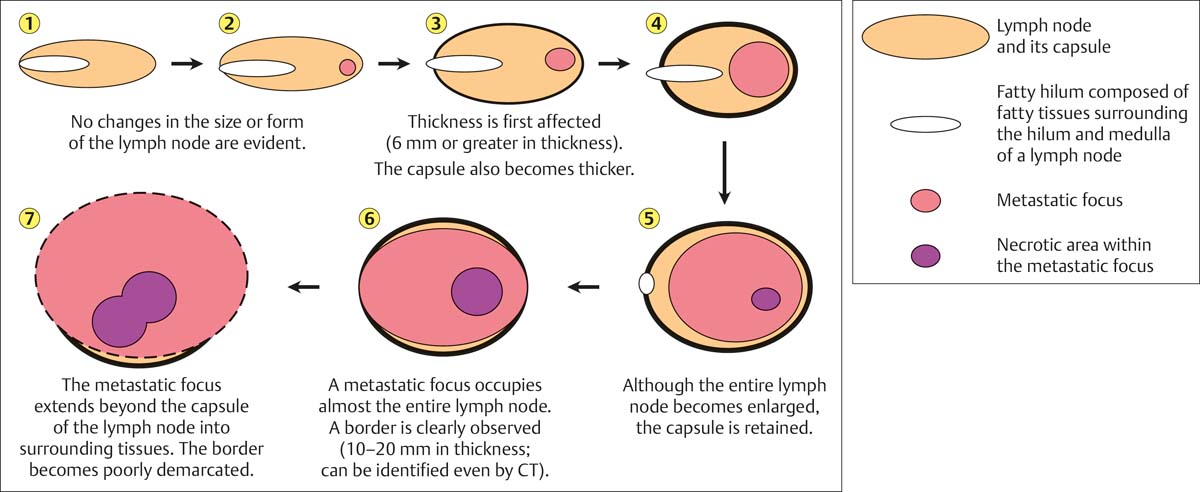
Neck Lymph Nodes | Radiology Key

Terms, definitions and measurements to describe sonographic features of lymph  nodes: consensus opinion from the Vulvar International Tumor Analysis  (VITA) group - Fischerova - 2021 - Ultrasound in Obstetrics & Gynecology -  Wiley Online Library
Terms, definitions and measurements to describe sonographic features of lymph nodes: consensus opinion from the Vulvar International Tumor Analysis (VITA) group - Fischerova - 2021 - Ultrasound in Obstetrics & Gynecology - Wiley Online Library

The procedure for lymph node measurement. The maximal-sized axis is... |  Download Scientific Diagram
The procedure for lymph node measurement. The maximal-sized axis is... | Download Scientific Diagram
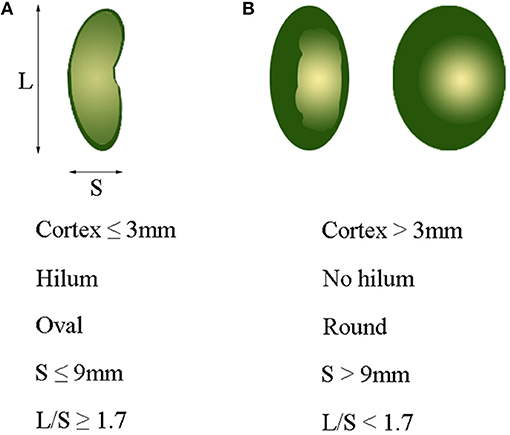
Frontiers | Predictive Value of Preoperative Multidetector-Row Computed  Tomography for Axillary Lymph Nodes Metastasis in Patients With Breast  Cancer
Frontiers | Predictive Value of Preoperative Multidetector-Row Computed Tomography for Axillary Lymph Nodes Metastasis in Patients With Breast Cancer
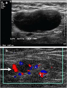
Lymphadenopathy - Wikiwand

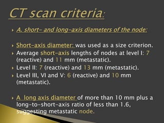
The Radiology Assistant : RECIST 1.1 - the basics

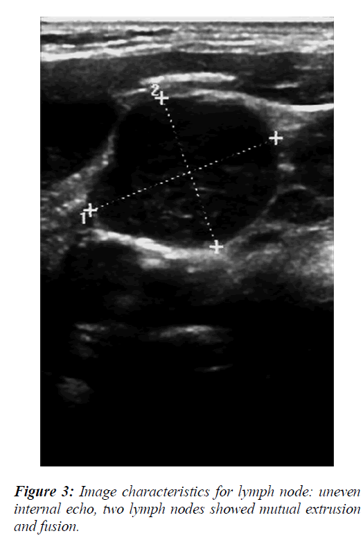
The logistic regression analysis of ultrasonographic features in  differential diagnosis of cervical lymph nodes metastasis of nasopharyngeal  carcinoma patients.
The logistic regression analysis of ultrasonographic features in differential diagnosis of cervical lymph nodes metastasis of nasopharyngeal carcinoma patients.
Drawing shows short axis of cortex (white arrow) and short axis of... |  Download Scientific Diagram
Drawing shows short axis of cortex (white arrow) and short axis of... | Download Scientific Diagram

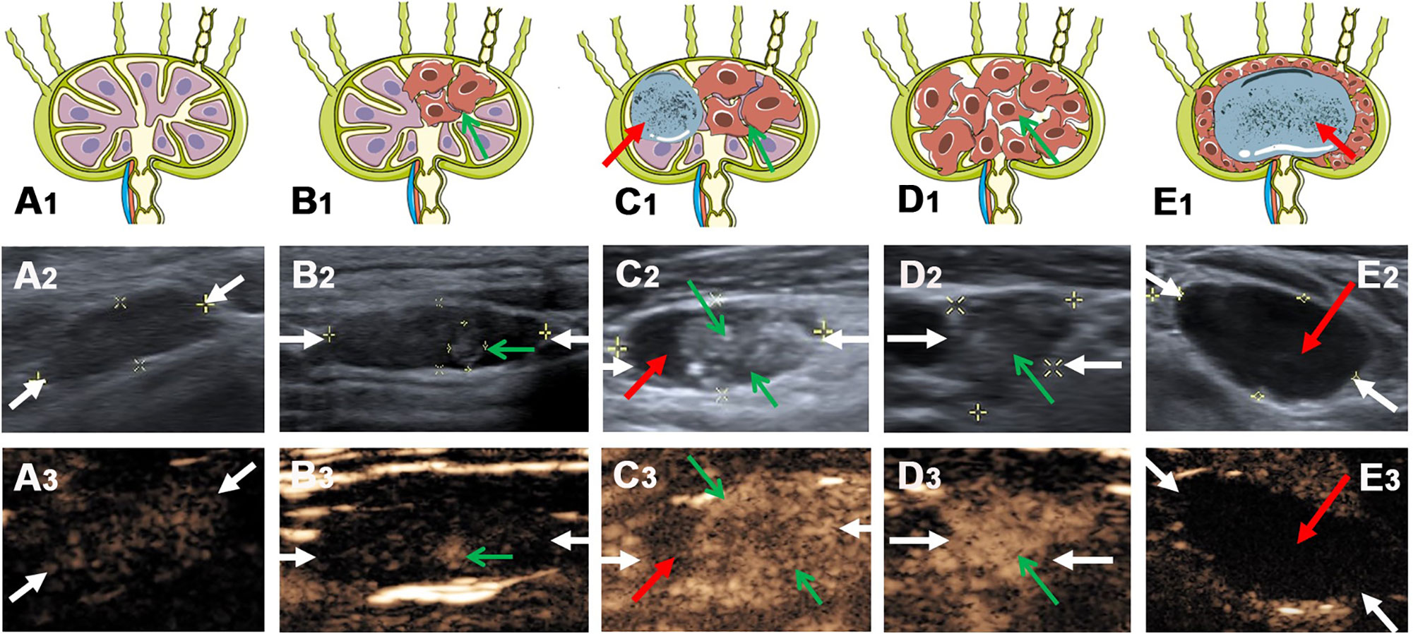
Frontiers | Value of Contrast-Enhanced Ultrasound for Evaluation of  Cervical Lymph Node Metastasis in Papillary Thyroid Carcinoma
Frontiers | Value of Contrast-Enhanced Ultrasound for Evaluation of Cervical Lymph Node Metastasis in Papillary Thyroid Carcinoma

Lymphadenopathy - Wikipedia

B-Mode and Elastosonographic Evaluation to Determine the Reference  Elastosonography Values for Cervical Lymph Nodes
B-Mode and Elastosonographic Evaluation to Determine the Reference Elastosonography Values for Cervical Lymph Nodes
Neck Lymph Nodes | Radiology Key

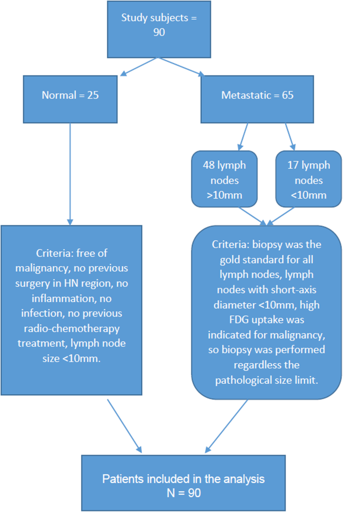
Diffusion-Weighted Imaging (DWI) derived from PET/MRI for lymph node  assessment in patients with Head and Neck Squamous Cell Carcinoma (HNSCC) |  Cancer Imaging | Full Text
Diffusion-Weighted Imaging (DWI) derived from PET/MRI for lymph node assessment in patients with Head and Neck Squamous Cell Carcinoma (HNSCC) | Cancer Imaging | Full Text

Contribution of Doppler Sonography Blood Flow Information to the Diagnosis  of Metastatic Cervical Nodes in Patients with Head and Neck Cancer:  Assessment in Relation to Anatomic Levels of the Neck | American
Contribution of Doppler Sonography Blood Flow Information to the Diagnosis of Metastatic Cervical Nodes in Patients with Head and Neck Cancer: Assessment in Relation to Anatomic Levels of the Neck | American

Figure 2 from Lymph Node Sonography | Semantic Scholar

Imaging of enlarged lymph node